

Medically reviewed by: Kinga Wojtczak, Doctor of Veterinary Medicine, specialist in canine and feline diseases
Cushing's disease in dogs is a chronic condition that seriously affects the animal's whole body. If you notice that your dog has been drinking a lot lately, has had a ravenous appetite for some time, and at the same time is weak and lacks energy, it is time to see a veterinarian. These signs occur in most patients with hyperadrenocorticism (an overactive adrenal cortex), but additional tests are needed to confirm the diagnosis.
In this article, you will learn which dogs are most at risk of Cushing's syndrome and how animals with this condition are treated. I will also explain what the diet of an affected dog should look like and what the consequences of ignoring the signs of Cushing's disease in dogs can be.
Table of contents:
What is Cushing's disease in dogs?

Overactivity of the adrenal cortex in dogs (hyperadrenocorticism / HAC), also called Cushing's disease or Cushing's syndrome, is a multisystem hormonal disorder in which the adrenal glands produce too much cortisol, which then acts on the body over a long period. Other names for this disease are hypercortisolemia and hyperadrenocorticism.
Hypercortisolemia results from:
- Abnormal function of the pituitary gland – this is called the pituitary form of Cushing's disease.
- Overactivity of the adrenal cortex caused by abnormal function of the adrenal glands – this is the adrenal form of Cushing's syndrome.
- Human action (administration of exogenous glucocorticosteroids) – this is called the iatrogenic form of Cushing's syndrome.
The terminology used for this condition is quite often confusing – some call it Cushing's disease, others use the name Cushing's syndrome. However, you should know that these terms do not mean the same thing. The main cause of the hypercortisolemia plays an important role here. So the term Cushing's syndrome is used when an adrenal disease is responsible for the abnormal glucocorticosteroid level, and Cushing's disease when the hypercortisolemia is caused by abnormal pituitary function.
Depending on the cause, the following forms are recognized:
- The pituitary form of Cushing's disease.
- The adrenal form of Cushing's syndrome.
- The iatrogenic form of Cushing's syndrome.
To better understand how the hormones work and how they affect the body's cells and tissues, let's look at three important structures that make up the hypothalamic–adrenal axis. They are:
Adrenal glands
- Adrenal glands – a pair of endocrine glands sitting on the upper poles of the kidneys (hence the name – they lie just above the kidneys). Each gland consists of the adrenal cortex and the adrenal medulla. * The adrenal cortex secretes steroid hormones essential for life, which can be divided into 3 main types: * Mineralocorticosteroids (aldosterone), * Glucocorticosteroids (cortisol, corticosterone, cortisone), * Androgens. * The adrenal medulla produces catecholamines: * Dopamine, * Norepinephrine (noradrenaline), * Epinephrine (adrenaline).
Effects of glucocorticosteroids
In Cushing's syndrome, cortisol plays the most important role, so let's take a closer look at its effects. They are numerous and very diverse. Cortisol is considered the stress hormone, and it is credited with the "fight-or-flight" response. Cortisol is a very necessary chemical compound, especially in situations that require an immediate reaction, but its long-term action on the body is devastating.
Put most simply, the main physiological role of cortisol is its broad effect on metabolism. The best-known metabolic effect is a very strong (sometimes even 5–10-fold) increase in gluconeogenesis. In this way, glucocorticosteroids maintain an adequate fasting blood glucose concentration and allow rapid energy production when the body is exposed to a threat.
This makes survival possible. Unfortunately, when cortisol acts for a long time, an "energy surplus" builds up that is not used and is diverted into abnormal side pathways (responsible for the signs of the disease). The metabolism of simple sugars is disturbed in two ways: on the one hand, their production increases (hepatic gluconeogenesis stimulated by cortisol), and on the other, their peripheral use is limited (glucocorticosteroids reduce glucose uptake by cells).
As a result, there is a strong tendency toward hyperglycemia (an increased blood glucose concentration), which leads to the development of diabetes of adrenal origin. This is known as the diabetogenic effect of cortisol. Cortisol increases protein catabolism and the mobilization of amino acids in tissues, especially in muscle and bone. Protein is the second (after glucose) source of energy that is drawn on in threatening situations; it is also an important building material for repairing any damage or wounds.
Many compounds formed by protein catabolism are starting materials for the gluconeogenesis mentioned earlier. It is no wonder, then, that increased protein breakdown under long-term steroid action leads to disorders of the skin (atrophy, poor wound healing, alopecia), bones (osteoporosis), and muscles and fasciae (a soft, flaccid abdominal wall). Cortisol has lipolytic and ketogenic effects. This means that it causes the mobilization of fatty acids from adipose tissue and increases their burning, thereby raising the concentration of free fatty acids in the blood.
Ketone bodies are yet another source of energy (after glucose and proteins). A characteristic feature of steroid action is that fatty acids, rather than glucose, are increasingly used to produce energy. This may lead to an increase in ketone body concentrations (the ketogenic effect of cortisol). As a result of long-term cortisol action, the total body fat content increases (despite the lipolytic effect) at the expense of protein, and fat distribution changes, with fat accumulating mainly in the dorsal part of the trunk.
In the limbs, on the other hand, muscle wasting occurs (the characteristic silhouette of an affected dog – a "ball of fat on four matchsticks"). Cortisol blocks the action of sex hormones. As a result, female dogs develop a prolonged anestrus (sexual resting phase) (delayed heat), and males develop testicular atrophy. Cortisol increases glomerular filtration in the kidneys and reduces the release of antidiuretic hormone (vasopressin).
The effect is increased diuresis. Clinically, this is seen as polyuria (increased urination). Cortisol potentiates the effects of epinephrine and norepinephrine on blood vessels by increasing their contractile reactivity (glucocorticosteroids in effect sensitize cells to catecholamines). In this way, they stimulate the contractility of the heart muscle and raise blood pressure.
Cortisol increases the release of gastrin in the stomach, which in turn leads to increased hydrochloric acid secretion. In the skeletal system, glucocorticosteroids cause demineralization and osteoporosis, while reducing the absorption of calcium ions and the reabsorption of phosphates from the intestines. The mobilization of calcium from bone in a stressful situation is related to the muscles' demand for this element.
However, dogs with excess cortisol may develop calcinosis cutis (abnormal foci of calcification in the skin); this is dystrophic calcification, which forms while the blood calcium concentration is normal. Cortisol and other glucocorticosteroids have strong anti-inflammatory effects and weaken the immune system. They reduce the body's hypersensitivity to antigens because they inhibit the release of histamine (this is called the cortisol block).
In this way, they prevent most life-threatening allergic reactions, such as anaphylactic shock or serum sickness. With long-term action, they cause atrophy of lymphoid tissue throughout the body, which reduces the production of immunoglobulins. This leads to weakened immunity. Excessive hormonal activity of the adrenal cortex, and with it a permanently raised blood cortisol level, leads to the development of a complex of multiple signs that together make up the picture of Cushing's syndrome.
Pituitary gland
The pituitary gland is a small gland that lies in a depression of the sphenoid bone – the sella turcica – at the base of the brain. It secretes a number of different hormones, of which the most important for us is adrenocorticotropic hormone – ACTH (also called corticotropin). This hormone stimulates the activity of the adrenal cortex, that is, it stimulates it to synthesize and release glucocorticosteroids.
Hypothalamus
The hypothalamus produces a hormone that releases corticotropin (CRH – corticotropin-releasing hormone; also called corticoliberin) in response to a stressful situation (e.g., a visit to the veterinarian or meeting a new dog). The job of CRH is to "inform" the pituitary gland that it needs to produce adrenocorticotropic hormone (ACTH). This hormone in turn travels to the adrenal glands, stimulating them to release cortisol.
Why? This stress hormone is meant to "rev up" the metabolism and provide anti-inflammatory and immunosuppressive effects. Such action is very beneficial and, if everything runs normally, highly desirable. However, at some point the secretion of hormones from the hypothalamus and the pituitary gland needs to be shut down so that the body can return to a state of rest. How does this blocking of secretion work under normal conditions?
At some point, high levels of circulating cortisol become a signal for the hypothalamus to stop producing CRH, which is meant to prevent the cycle from starting again. This is called "negative feedback," and it is a very important mechanism that checks, controls and keeps the body in balance by counteracting excessive cortisol release. However, this self-regulating cycle of releasing and inhibiting hormone production is much more complex.
This is because cortisol has an inhibitory effect on both the pituitary gland and the hypothalamus – that is, there is both secondary (pituitary) and tertiary (hypothalamic) negative feedback. The problem of excessive cortisol production by the adrenal glands may therefore originate in the adrenal gland itself (this is called primary hyperadrenocorticism) or in the pituitary gland, when ACTH is overproduced (this is referred to as secondary hyperadrenocorticism).
In dogs, tertiary hyperadrenocorticism is also probable, associated with a hypothalamic disorder and excessive production of corticoliberin (CRH).
Pituitary-dependent Cushing's syndrome
Pituitary-dependent Cushing's syndrome (Pituitary-Dependent Hyperadrenocorticism – PDH) is the predominant form of Cushing's syndrome, accounting for about 80–90% of cases of spontaneous hypercortisolemia, and is usually caused by a pituitary tumor (an adenoma or an adenocarcinoma).
The most commonly recorded pituitary tumors are:
- adenomas of the pars distalis,
- less often, adenomas of the pars intermedia,
- very rarely, pituitary carcinomas.
PDH is characterized by abnormally elevated levels of cortisol and ACTH. Put simply, pituitary disease dysregulates the hypothalamic–pituitary–adrenal axis and makes ACTH production somewhat "erratic" (despite the elevated blood cortisol level). As a result, the pituitary gland releases excessive amounts of corticotropin, both in terms of frequency and intensity.
The causes of PDH are related to various pituitary tumors. We distinguish:
- Pituitary Cushing's disease associated with pituitary macrotumors.
Macroadenomas account for 10–20% of cases. These are large pituitary tumors over 1 cm that tend to extend beyond the upper border of the gland, reaching the hypothalamus. The signs seen with these tumors may be somewhat different from those typical of Cushing's syndrome. Various ophthalmic signs often appear, as well as changes in the dog's behavior: * aggression, * sensory disturbances, * appetite disturbances. - Pituitary hyperadrenocorticism associated with pituitary microtumors (most cases).
Most pituitary tumors are chromophobe microadenomas. They are usually well-circumscribed, clearly localized tumors of small size (less than 1 cm in diameter).
Regardless of the cause, however, excessive ACTH secretion by the pituitary gland stimulates the adrenal cortex, increasing cortisol secretion and leading to bilateral hyperplasia of the cortex. With a pituitary adenoma, negative feedback becomes relatively ineffective – however, hypothalamic function and the action of CRH in controlling ACTH release are suppressed.
Surprisingly, in many cases ACTH and cortisol levels are, as a rule, within the reference ranges. The pituitary form of Cushing's disease most often affects middle-aged and older dogs (the average age is about 7–9 years). It can occur in all dog breeds, but dachshunds, poodles and small terriers are predisposed.
Adrenal-dependent Cushing's syndrome
The second type of Cushing's syndrome is caused by functional (cortisol-producing) adrenal tumors. These may be adenomas or carcinomas (which occur with equal frequency). Carcinomas are usually larger, may invade surrounding tissues and may metastasize to distant sites (mainly the liver and lungs). Adrenal tumors are responsible for about 15–20% of cases of hypercortisolemia. An adrenal tumor is usually unilateral.
It produces extra cortisol in an uncontrolled way, independently of the pituitary gland and ACTH. Put simply, it escapes control and, even without stimulation from the pituitary, produces excess cortisol, which in turn suppresses ACTH secretion by the pituitary gland. As a result, the healthy adrenal gland (not affected by the tumor) reduces its activity, which leads to atrophy of its cortex. In the gland in which the tumor develops, on the other hand, the normal cells gradually atrophy.
Because of the high cortisol level, CRH and ACTH secretion is inhibited (through negative feedback), but this does not matter in the case of adrenal tumors. Here, there are episodes of "random", entirely haphazard cortisol release by the adrenal tumor. Nevertheless, adrenal tumors may respond to ACTH stimulation. Note that there are also adrenal tumors that do not release cortisol.
These are usually adenomas that produce adrenal steroids other than cortisol. Their presence may cause signs very similar to Cushing's syndrome, but the blood cortisol level is low. Levels of other glucocorticosteroids, however, are high. Adrenal tumors appear more often in dogs of larger breeds; about 50% of patients weigh > 20 kg. The pituitary and adrenal forms of Cushing's syndrome are classified as idiopathic Cushing's syndrome.
The form caused by the administration of exogenous steroids is classified separately; it is called iatrogenic Cushing's syndrome.
Which dogs get Cushing's syndrome
Hypercortisolemia (excess cortisol) most often develops in middle-aged dogs (around 6 years old), although there are also cases in which the disease affects one-year-old dogs.
It is diagnosed in many dog breeds, but some breeds are strongly predisposed to it:
- Poodles,
- dachshunds,
- terriers (Yorkshire terrier, Jack Russell terrier, Staffordshire bull terrier, fox terrier),
- German shepherds,
- beagles,
- Labradors,
- as well as boxers and Boston terriers.
About 55–65% of cases are females. Cushing's syndrome affects dogs older than 9 years, with the average falling in the 11th year of life. The pituitary form of Cushing's disease appears to occur more often in small dogs (75% of cases weigh less than 20 kg), whereas about 50% of dogs with functional adrenal tumors weigh more than 20 kg.
Iatrogenic Cushing's syndrome in dogs
Iatrogenic hypercortisolemia is caused by long-term, uncontrolled use of glucocorticoids (including topical use, e.g., in eye drops, ear drops, etc.). Chronic administration of exogenous steroids suppresses ACTH secretion by the pituitary gland and leads to atrophy of the zona fasciculata of the adrenal cortex and a drop in the production of endogenous glucocorticoids. Iatrogenic Cushing's syndrome occurs regardless of the animal's age, breed or sex.
Cushing's disease in dogs: symptoms
Hyperadrenocorticism produces one of the widest ranges of clinical signs of any condition in veterinary medicine. The multitude and variety of signs and their varying severity result from the multi-organ effects of adrenocortical hormones. Cushing's syndrome is a systemic disease in which some signs are typical and appear in most patients, while others may be milder or not appear at all.
Many of them are hard for the owner to notice and develop over months or even years. They may also gradually worsen, or periodically subside and return. The more signs occur together, the more likely hypercortisolemia is.
General signs
Polyuria and polydipsia
Polyuria and polydipsia (PU/PD) – increased urination and increased thirst (80–85% of cases). This is the most common reason for a visit to the veterinary clinic and probably the most specific sign in patients with hypercortisolemia.
In over 85% of dogs with hyperadrenocorticism, it appears as the first sign:
a worried owner reports that the dog cannot last the whole night without urinating.
Polyuria results from the action of cortisol:
on the one hand, it reduces the release of antidiuretic hormone; on the other, it increases glomerular filtration in the kidneys. This leads to a significant increase in the frequency of urination. Urinary incontinence in dogs also often appears. Polyuria is the primary sign here, while increased water intake appears secondarily, to compensate for the losses. If a dog drinks more than 60 ml/kg body weight of water a day, this is called polydipsia.
The increase in thirst is comparable to that seen in diabetes – the dog's water intake is increased 2- to 6-fold and may reach 100 ml/kg body weight/day or even more.
Polyphagia
Polyphagia – increased appetite (>90% of cases). Another very common sign that owners report to the veterinarian. At first it may suggest diabetes; in fact, it is very likely that the increased appetite results from reduced insulin activity. Polyphagia is also caused by the neurological action of cortisol.
Weakness, lethargy, sleepiness
Quite often a dog with Cushing's syndrome becomes lethargic. It is not the first sign reported, but it occurs as often as those listed above.
Heavy panting and puffing
Change in body shape
A change in body shape occurs in over 80% of cases.
- Very characteristic of Cushing's syndrome is the Cushingoid body shape: * an enlarged, pendulous abdomen (associated with liver enlargement, muscle weakness and fat accumulation in the abdominal cavity), * uneven fat distribution (mainly on the trunk), * muscle wasting in the limbs.
The Cushingoid body shape results from the action of glucocorticosteroids, which increase the tendency to fat deposition (fat metabolism is disturbed, and fat tends to accumulate along the dorsal midline) and to slackening of the musculofascial structures (reduced abdominal wall tone, abduction of the limbs, in which muscle mass gradually wastes away). As a result, the dog looks like "a ball on four matchsticks".
Liver enlargement
In hypercortisolemia, the liver enlarges and "rests" on the already slack abdominal wall, which stretches it even further. Hepatomegaly results from the action of steroids, and this complex liver overload is called steroid hepatopathy.
Neurological and muscular signs
Neurological and muscular signs occur in 75–85% of dogs. A fairly commonly reported problem is lethargy and muscle weakness. The dog looks as if it has no strength.
In dogs with Cushing's disease, myopathy may develop, characterized by stiffness of the limbs (mainly the hind limbs). This causes problems with movement:
the dog moves with a hopping gait, often without bending its knees. This disorder is not painful, but as it progresses, it leads to severe muscle wasting, and over time the animal becomes increasingly helpless and infirm. Neurological signs that are not uncommon also include facial paralysis and pseudomyotonia.
- Heat intolerance (risk of overheating)
- Abdominal obesity.
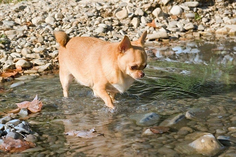
Abdominal obesity appears along with the increase in appetite, and owners often regard it as a sign of good health.
Skin signs
Dermatological signs are very common and usually strongly suggest Cushing's disease (together with PU/PD). Skin changes are often the first sign the owner reports.
Hair loss (alopecia)
It may appear as the first sign and at first not raise suspicion of hypercortisolemia. However, this sign may be absent, and the absence of hair loss does not rule out hyperadrenocorticism. It is caused by the effect of cortisol on the anagen (growth) phase of the hair cycle. The remaining coat is dominated by dull, matted hairs that may also be discolored (this is the telogen phase of hair growth).
If the hair loss is generalized, bilateral and symmetrical, its location may be specific to the given dog breed. The typical location is the trunk, but sometimes it occurs only on the bridge of the nose (linear alopecia) or as a rat tail. In poodles, hair loss is usually the first worrying sign. It first appears on the sides of the body and at the base of the limbs, while the head and the peripheral parts of the body stay covered with hair longer.
In dachshunds, hair loss is more diffuse. Typical sites are the abdomen, the outer surface of the ear flaps, the base of the tail, but also the sides of the body and the limbs.
Thin skin with reduced elasticity
Normally, a dog's skin is thickest on the dorsal neck and trunk and thinnest on the limbs, especially on the digits. In Cushing's syndrome, the skin becomes thin, especially in the places where the changes are greatest.
This is usually the abdominal area, where the umbilical veins are very clearly visible through the skin, and the inner thighs:
the skin there is as thin as tissue paper. This results from the atrophy of the skin's connective tissue under the influence of cortisol. In addition, the skin loses elasticity. When lifted into a fold, it slowly returns to its original position (normally, a skin fold flattens immediately after it is released). The skin wrinkles, as if there were too much of it for the size of the body – this is clearly visible as numerous folds, especially around the hocks.
Pigmented spots
Melanin-containing pigmented spots (hyperpigmentation) are particularly common. They are scattered all over the animal's body. The spots are not specific to Cushing's syndrome, but their presence may strengthen the suspicion of this disease.
- A tendency to develop phlebectasias (small dilated veins in the skin), visible as small, blood-red streaks.
- A tendency to bruise
- Skin stretch marks (striae) that form spontaneously or at scar sites. At the sites of previous surgeries, scars may come apart.
- Comedones (blackheads).
Black seborrheic bumps on the skin of the abdomen, especially around the mammary glands and in the perianal area, are very characteristic of hyperadrenocorticism. They additionally predispose the dog to secondary hair follicle infections.
Calcinosis cutis
It consists of deposits of insoluble calcium salts in the skin and subcutaneous tissue (mineralization). The lesions are most often located on the back, especially on the neck and in the sacral area, in the armpits and in the groin. Generalized calcinosis cutis accompanying hypercortisolemia is dystrophic calcification; the blood calcium concentration is usually normal. It can take various forms – from very fine deposits of calcium salts visible only under a microscope, through single small nodules, to large yellowish deposits that are raised and clearly demarcated from the neighboring tissues.
If calcinosis is accompanied by secondary infections, there is also itching and pain, as well as scabs located in the affected areas of the back, neck and nose.
Poor wound healing
It can cause chronic ulceration and results from reduced fibroblast proliferation and decreased collagen synthesis.
Secondary skin diseases
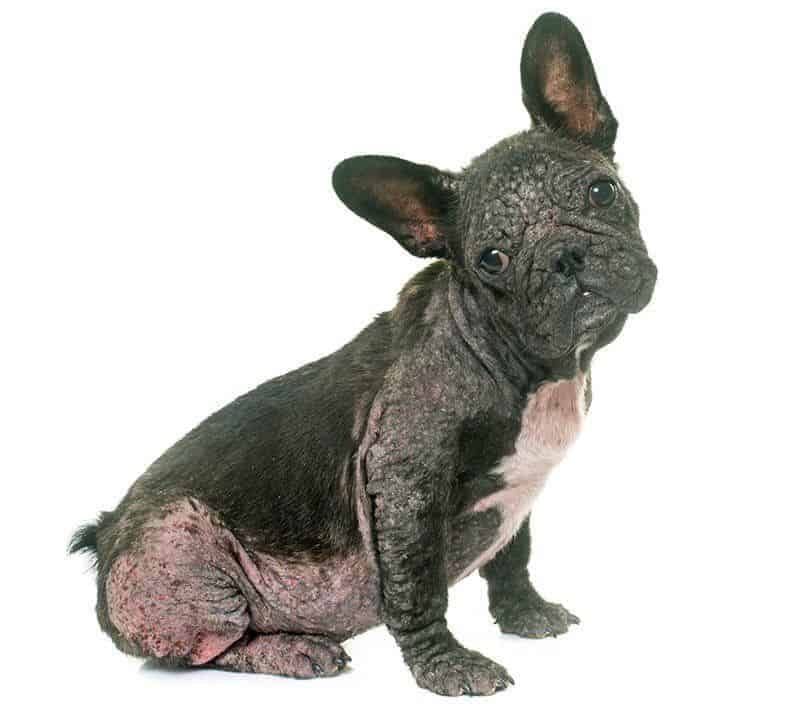
- Many skin diseases can complicate the course of Cushing's syndrome: * seborrhea, * bacterial infections, * fungal infections, * Demodex mite infestations.
The response to treatment in such cases is poor, especially if the hyperadrenocorticism is not treated at the same time.
Reproductive problems
You may see:
- absent or delayed heat,
- testicular atrophy,
- an enlarged clitoris, resulting from overproduction of adrenal androgens.
Neurological signs
Neurological signs associated with a pituitary macroadenoma:
- lethargy,
- dullness,
- decreased appetite,
- wandering, aimless roaming,
- circling, head pressing,
- behavioral changes,
- Horner's syndrome,
- seizures (rare).
Signs and conditions not specific to Cushing's syndrome
These may also be present:
- absent or reduced appetite,
- vomiting,
- diarrhea,
- sneezing,
- coughing,
- jaundice,
- itching,
- pain,
- lameness caused by inflammation,
- epileptic seizures,
- bleeding,
- kidney failure,
- pancreatitis,
- liver failure,
- immune-mediated diseases,
- ligament ruptures,
- eye signs – bulging eyes (exophthalmos), corneal ulcers, blindness.

Cushing's syndrome: complications
Complications develop in some animals; they include:
- high blood pressure (systemic hypertension; present in about 50% of cases),
- urinary tract infections,
- pyelonephritis (kidney infection),
- bladder inflammation (cystitis; clinically silent),
- urinary stones (urolithiasis),
- calcium-containing stones,
- struvite stones, if associated with infections,
- congestive heart failure,
- pancreatitis,
- diabetes,
- impaired wound healing,
- recurrent infections,
- joint laxity,
- hypercoagulability,
- pulmonary thromboembolism,
- aortic thromboembolism.
Cushing's syndrome: diagnosis
Cushing's syndrome is often a very frustrating condition to diagnose, because many of the signs – even typical ones – may be absent, laboratory results can be ambiguous, and concurrent conditions can obscure the typical clinical signs. Affected dogs are usually not in a life-threatening state. The disease usually progresses slowly, and most patients show only one or a few of the typical signs.
Cushing's syndrome is a clinical syndrome that cannot be diagnosed without typical clinical signs. That is why the diagnostic process can be tedious and lengthy.
Cushing's syndrome should be suspected when the following abnormalities are present:
Always when:
- The dog has polyuria and polydipsia (PU/PD).
- There is symmetrical, bilateral hair loss.
- The dog has a "Cushingoid silhouette" (abdominal obesity and weakened abdominal wall tone).
- With these signs – after ruling out other diseases, of course (e.g., diabetes, hypothyroidism, kidney failure, etc.) – Cushing's syndrome should be included in the differential diagnosis.
Possibly when:
- There is hyperpigmentation (discoloration) of the skin.
- There is calcinosis cutis.
Taking a detailed history (including information about any steroids given) is always a very important part of the diagnosis. Once hypercortisolemia is suspected, a thorough laboratory workup should be performed.
Complete blood count
- In Cushing's syndrome, a fairly typical white blood cell pattern very often develops: the stress leukogram. This pattern is seen in almost all animals: * Leukocytosis with neutrophilia, * Monocytosis, * Lymphopenia, * Eosinopenia.
- Similarly, thrombocytosis – an increased platelet count – affects almost all dogs with hypercortisolemia.
- Some dogs may have mild erythrocytosis – an increased red blood cell count.
Serum biochemistry
- Elevated blood glucose – hyperglycemia.
Most often this is a moderate rise in blood sugar and – in most cases – without glucose in the urine (glucosuria) at the same time. In about 50% of cases it is accompanied by a simultaneous rise in insulin levels. This is the effect of intense gluconeogenesis and reduced peripheral glucose use due to insulin antagonism. Cushing's disease should always be considered in a diabetic dog in which it is difficult to establish an effective insulin dose.
Hypercortisolemia causes insulin resistance and may lead to the development of diabetes. Interestingly, dogs with concurrent diabetes do not show the typical signs of excessive cortisol. Even so, Cushing's should always be considered if normoglycemia cannot be achieved in a diabetic dog.
- Increased alkaline phosphatase (ALP) activity (>150 U/L, sometimes > 1000 U/L) occurs in 90–95% of dogs with hypercortisolemia.
The rise in alkaline phosphatase is marked (usually about 5–10 times above the reference range). It is considered a consistent, though non-specific, criterion in diagnosing Cushing's disease.
- Increased alanine aminotransferase (ALT) – found in over 90% of dogs with hypercortisolemia. It is caused by the liver being overloaded with glycogen.
- Elevated blood cholesterol (due to increased lipolysis).
In about 50% of patients this parameter is not very specific, but together with other characteristic abnormalities it may suggest Cushing's syndrome. Elevated cholesterol and elevated serum alkaline phosphatase are the most reliable indicators of hypercortisolemia at this stage of the diagnosis. In some patients, ALP may remain within the normal range.
- Blood urea nitrogen (BUN) is usually normal or may be decreased.
- Elevated triglycerides (as a result of cortisol-induced lipolysis).
- Slightly elevated fatty acids.
- Slightly elevated sodium.
- Slightly decreased potassium.
Urinalysis
- Urine specific gravity < 1.020, often even <1.008. In about 50% of patients, the ability to concentrate urine is impaired or absent (hypo- or isosthenuria).
- Proteinuria (protein in the urine).
- Bacterial urinary tract infection – in 40–50% of cases: * the infection is usually occult, so a bacterial culture of a urine sample collected by cystocentesis is necessary, * it results from incomplete emptying of the bladder caused by muscle weakness, as well as from immunosuppression related to the action of glucocorticoids.
- A slight increase in the urine protein:creatinine ratio (UPC) – usually less than 5.
Survey abdominal radiographs
- liver enlargement (hepatomegaly),
- a fairly large amount of abdominal fat, which provides good radiographic contrast,
- an overdistended bladder,
- urinary stones (urolithiasis),
- calcification (mineralization) of soft tissues,
- osteoporotic changes in the vertebrae,
- adrenal calcification – rare; usually associated with an adrenal adenoma or carcinoma.
Survey chest radiographs
- calcification in the airways,
- osteoporotic changes in the vertebrae,
- lung metastases (rare),
- pulmonary thromboembolism.
Abdominal ultrasound
- enlarged adrenal glands (in the pituitary form of Cushing's syndrome),
- an adrenal tumor with a simultaneous reduction in the size of the other gland (in the adrenal form of Cushing's syndrome),
- calcification within the adrenal gland (with a tumor),
- metastases,
- liver enlargement (hepatomegaly),
- increased echogenicity of the liver,
- a distended, atonic bladder,
- urinary stones (urolithiasis),
- soft tissue calcification.
Advanced imaging
Both computed tomography and magnetic resonance imaging are useful in diagnosing adrenal tumors or adrenal hyperplasia and large pituitary tumors, but because of their fairly high cost they are still used too rarely.
- Computed tomography or a brain MRI may reveal a pituitary tumor * This test is recommended to confirm the cause of neurological signs, and also when radiation therapy or surgery is being considered. * Abdominal computed tomography is recommended before adrenalectomy (removal of the adrenal gland).
- Histopathology (in the case of tumors).
As the likelihood of Cushing's syndrome or disease increases, the next step is to test adrenal function.
Screening tests
The aim of hormone testing is to show increased cortisol production by the adrenal glands or reduced sensitivity of the hypothalamic-adrenal axis to the negative feedback of steroids. These tests are performed only when clinical signs of hypercortisolemia are present, because only then do they have diagnostic value.
Baseline blood cortisol
Measuring cortisol levels is the main part of the laboratory diagnosis of Cushing's syndrome. This test has rule-out value – if cortisol levels are normal, Cushing's syndrome is unlikely. However, if the level of this hormone is elevated, additional tests should be performed. This is because even in healthy but stressed animals, cortisol concentrations may exceed the reference range.
Cortisol concentrations fluctuate quite widely during the day, and baseline levels are usually higher with stress or other diseases. That is why a single cortisol measurement is of little use in making a diagnosis and should be supplemented with additional tests.
Urine cortisol:creatinine ratio (UC/C)
Dogs with Cushing's syndrome have elevated serum cortisol levels and excrete larger amounts of it in the urine. Because the amount of cortisol in the blood can rise with a physiological increase in metabolic rate, it is expressed relative to the urine creatinine value. Its concentration may vary between urine samples collected at different times of the day. It is a simple method, introduced because it is easy to perform and makes it possible to eliminate stress.
It is suitable mainly as a screening test. The owner collects the dog's morning urine sample on 2 consecutive days and keeps it in the refrigerator. The ratio of free urinary cortisol to creatinine is then calculated.
Urine test protocol:
- Collect the morning urine on the first day – the first sample.
- Collect the morning urine on the second day – the second sample.
- Sometimes (to differentiate pituitary from adrenal Cushing's syndrome): * Oral dexamethasone on the second day at a dose of 0.1 mg/kg body weight three times a day, * Collect the morning urine on the third day.
Interpreting the urine test:
- Urine cortisol (nmol/L) / urine creatinine (mmol/L) ×10⁻⁶: * Ratio: * Day 1: <15×10⁻⁶ – negative result. This is not Cushing's syndrome * Day 1: between 15×10⁻⁶ and 25×10⁻⁶ – equivocal result * Day 1: >25×10⁻⁶ – Cushing's syndrome in >93% of cases * With two measurements: the mean of the ratios from the first 2 days is calculated and assessed as above. * If the ratio is elevated in samples 1 and 2 – assess the UC/C with the suppression test. Ratio: * Day 3. A UC/C ratio >50% of the mean of the first two samples may indicate the adrenal form, although the pituitary form resistant to dexamethasone suppression is also not ruled out. * Day 3. A UC/C ratio <50% of the mean of the first two samples indicates the pituitary form of Cushing's syndrome.
In spontaneous Cushing's syndrome, the urinary cortisol level is higher than in healthy animals. The test is relatively sensitive but unfortunately not specific for hyperadrenocorticism, because similar values are found with other causes of polyuria and polydipsia. In addition, there are many false-negative results. Not all dogs with Cushing's syndrome have an abnormal concentration. It is a good screening test for dogs suspected of Cushing's syndrome that do not have severe clinical signs.
So measuring the urine cortisol/creatinine ratio makes it possible to rule out hypercortisolemia when the result is normal. Unfortunately, an elevated UC/C ratio does not allow a definitive diagnosis. Abnormal urine cortisol/creatinine ratios may be found in other conditions, such as diabetes mellitus, diabetes insipidus, pyometra, hypercalcemia, kidney disease and liver disease. That is why this test is better used to monitor treatment than to establish the diagnosis.
Nor does this method differentiate PDH from ADH, unless the cortisol/creatinine ratio is 100×10⁻⁶, which suggests PDH.
ACTH stimulation test
It consists of measuring the dog's cortisol level before and after stimulating the adrenal glands with an injection of ACTH. This makes it possible to test the maximum secretory capacity of the adrenal cortex. The test relies on the fact that hyperplastic adrenal glands can produce more cortisol when they are stimulated. It also makes it possible to show hypocortisolemia caused by corticosteroid therapy (suppression of adrenal function).
This is very important information, because together with the clinical signs it makes it possible to determine whether the change is associated with iatrogenic Cushing's syndrome.
In dogs, the ACTH stimulation test is the method of choice for:
- diagnosing iatrogenic Cushing's syndrome,
- monitoring the treatment of hyperadrenocorticism,
- monitoring the patient after adrenalectomy,
- also for diagnosing hypoadrenocorticism (Addison's disease).
Test protocol (fasted dog):
- The first blood sample is taken to establish the baseline cortisol level. ACTH is injected intramuscularly or intravenously at a dose of 0.25 mg. In a healthy animal, the cortisol level before ACTH is given is 20 to 250 nmol/l (0.7–9 µg/dl).
- The second blood sample is taken one hour after the injection = the cortisol level after ACTH stimulation. In a healthy animal, the level should be between 200 and 450 nmol/l (9–16 µg/dl).
Interpreting the result:
- Cortisol level after ACTH stimulation: * <17 µg/dl (<470 nmol/l) – normal result * > 22 µg/dl (> 607 nmol/l) – elevated level – Cushing's disease very likely * 17–22 µg/dl (470–607 nmol/l) – equivocal result ("gray zone") – Cushing's disease likely, but additional tests should be performed and the patient's clinical signs and their severity taken into account in the diagnosis
- Cortisol level before and after ACTH stimulation: * Both values <0.5–2 µg/dl – iatrogenic Cushing's syndrome or hypoadrenocorticism (suspected Addison's disease).
The interpretation of results differs between laboratories, but usually there is a clear response to ACTH – concentrations after 1 hour are as a rule three times higher than baseline levels. However, regardless of the baseline cortisol concentration, when its level after ACTH administration is above 600 nmol/l, hyperadrenocorticism should be diagnosed. This test has the highest specificity but low sensitivity.
There are many false-negative results, so a positive result gives the greatest certainty. Because dogs with the iatrogenic form of Cushing's syndrome show a greatly reduced response to ACTH stimulation, it is the test of choice when this condition is suspected. However, it cannot distinguish between the possible causes of Cushing's syndrome and should be supplemented with another test (a suppression test). In addition, some dogs with adrenal tumors respond normally to stimulation.
Low-dose dexamethasone suppression test (LDDST) – fasted patient!
The purpose of this test is to assess the pituitary-adrenal axis, which in spontaneous Cushing's syndrome is abnormally resistant to suppression by exogenous steroids (dexamethasone). It is a more sensitive method than the ACTH stimulation test, but less specific (false-positive results are more common). However, it gives high certainty when the result is negative. An advantage of this test is that it can differentiate the cause of Cushing's syndrome and determine whether we are dealing with the pituitary or the adrenal form of the disease.
Unfortunately, it is not a useful method for diagnosing iatrogenic Cushing's syndrome or hypoadrenocorticism. The test consists of measuring the cortisol level before and after an injection of a direct-acting glucocorticoid. Dexamethasone is generally used for this. The normal response to an exogenous steroid is a drop in the cortisol level within a few hours, because dexamethasone suppresses its production (through negative feedback it reduces ACTH secretion).
After about 2–3 hours, ACTH secretion is suppressed, and this lasts about 24–48 hours. The adrenal cortex produces less cortisol – its blood level falls. In Cushing's syndrome this suppression is much weaker, especially with adrenal tumors that produce cortisol autonomously (independently of ACTH). Giving dexamethasone will affect ACTH secretion by suppressing it, but it will not reduce cortisol secretion. As a result, the cortisol level will not fall or will drop only slightly.
Here the pituitary gland will respond, but the adrenal glands will not. In the pituitary form of Cushing's disease, dexamethasone has very little effect on the pituitary gland (or none at all). After the steroid injection, ACTH secretion is either not stopped at all or suppressed only briefly, after which the pituitary releases the hormone again and stimulates the adrenal cortex to produce cortisol.
The effect:
the cortisol level falls only very slightly or not at all, and drops only for a short time. This test is not suitable for diagnosing iatrogenic Cushing's syndrome or for monitoring the course of treatment. In practice, the doses of dexamethasone used have changed.
Two tests are currently performed:
suppression with low (0.01–0.02 mg/kg body weight) and high (0.1–1 mg/kg body weight) doses of dexamethasone.
Test protocol:
- First blood sample – baseline cortisol level (hour 0).
- Dexamethasone injected intravenously at a dose of 0.01 mg/kg body weight.
- Second blood sample 4 hours after the steroid injection (hour 4). It gives an indication of the deposition of residues and allows the form of Cushing's syndrome to be finally determined: pituitary or adrenal.
- Third blood sample 8 hours after the injection (hour 8) – measurement of the suppressed cortisol values.
Interpreting the test:
When interpreting the test results, the cortisol concentration in the sample taken 8 hours after the dexamethasone injection is considered first.
- If the result is within the reference range, then: * either the dog does not have hyperadrenocorticism, * or (with a probability of about 5–10%) it has pituitary-dependent hyperadrenocorticism. A dog may have an early form of pituitary disease that still responds to a pharmacological dose of steroid with reduced ACTH secretion, which lowers the blood cortisol level. In such situations, an ACTH stimulation test should be performed.
- If the cortisol concentration is above the upper limit of the reference range, the dog most likely has hyperadrenocorticism (false-positive results are possible with diseases unrelated to the adrenal glands). The cortisol concentrations in the baseline sample and in the sample taken at hour four are then assessed to check how cortisol secretion was suppressed throughout the test: * if a drop in blood cortisol concentration of at least 50% is seen at hour four or eight, the final diagnosis is pituitary-dependent hyperadrenocorticism, and no further adrenal function tests are needed.
- Healthy patient: * hour 0 – cortisol 1–5 µg/dl – normal or slightly elevated values (due to stress), * hour 4 – cortisol <1.4 µg/dl (<50% of baseline values, or <10ng/ml), * hour 8 – cortisol <1.4 µg /dl / <40 nmol/l (further suppression after 8 hours <10ng/ml).
- Pituitary-dependent hyperadrenocorticism: * "suppression" occurs in 60–65% of dogs with PDH, * when the cortisol concentration in the 4-hour sample is <1.4 µg/dl and after 8 hours >1.4 µg/dl, * when the cortisol concentration in the 4-hour sample is <50% of the baseline value and after 8 hours > 1.4 µg/dl (> 40 nmol/l), * when the cortisol concentration after 8 hours is <50% of the baseline value but always > 1.4 µg/dl.
- Pituitary-dependent or adrenal-dependent hyperadrenocorticism: * when the cortisol level in both the 4-hour and the 8-hour sample is > 1.4 µg/dl, * a complete absence of any effect suggests secreting (functional) adrenal tumors.
Interpreting the results of the low-dose dexamethasone suppression test is often difficult. Here are a few examples:
Patient 1
Cortisol levels in the low-dose dexamethasone suppression test:
- hour 0 – 10 µg/dl,
- hour 4 – 10 µg/dl,
- hour 8 – 10 µg/dl.
Interpretation:
We are certainly dealing with hyperadrenocorticism, because the cortisol concentration at hour 8 after the steroid is given is > 1.4µg/dl. Because the cortisol level was abnormally high in all samples, the results strongly suggest adrenal-dependent Cushing's syndrome, since with adrenal tumors suppression is not seen or is very weak. However, AT (adrenal tumor) cannot be definitively diagnosed on the basis of such results, because as many as 30% of dogs with the pituitary form of the disease also do not respond with a drop in cortisol. That is why the next differentiating test should be measurement of endogenous ACTH.
Patient 2
Cortisol levels in the low-dose dexamethasone suppression test:
- hour 0 – 10 µg/dl,
- hour 4 – 6 µg/dl,
- hour 8 – 4 µg/dl.
The cortisol level at hour 8 is higher than 1.4 µg/dl – hyperadrenocorticism is present. Because cortisol secretion is never suppressed in the test in dogs with adrenal tumors, the adrenal form of the disease cannot be diagnosed here. 8 hours after the steroid was given, the cortisol level is 4µg/dl; there was at least 50 percent suppression of cortisol secretion, which allows a diagnosis of pituitary-dependent hyperadrenocorticism.
Patient 3
Cortisol levels in the low-dose dexamethasone suppression test:
- hour 0 – 10 µg/dl,
- hour 4 – 4 µg/dl,
- hour 8 – 10 µg/dl.
Once again, a cortisol level > 1.4 µg/dl indicates hyperadrenocorticism. Although the cortisol levels at hour 0 and hour 8 are the same, this is not the adrenal form of the disease, because there was at least 50% suppression of secretion during the test (level at hour 4: 4 µg/dl).
The diagnosis is:
pituitary-dependent hyperadrenocorticism. The rise in cortisol between hour 4 and hour 8 reflects the faster metabolism of dexamethasone during that time, which is often present in patients with hyperadrenocorticism.
Patient 4
Cortisol levels in the low-dose dexamethasone suppression test:
- hour 0 – 10 µg/dl,
- hour 4 – 6 µg/dl,
- hour 8 – 1 µg/dl.
Despite the high baseline cortisol level (10µg/dl), its concentration fell to the reference range after 8 hours (<1.4µg/dl). That is why such a result should be approached very cautiously. On the one hand, it does not point to hyperadrenocorticism, but keep in mind that as many as 10% of dogs with the pituitary form of the disease may have normal test results.
In such a situation – in patients with clinical signs of hyperadrenocorticism and normal suppression test results – an ACTH stimulation test should be performed, or you should wait 4–6 weeks and repeat the test.
High-dose dexamethasone suppression test (HDDST)
In the pituitary form of Cushing's syndrome, negative feedback is not completely abolished, whereas in the adrenal form cortisol secretion is not influenced by exogenous steroids. High doses of dexamethasone (0.1 mg/kg body weight) suppress the release of ACTH by the pituitary gland, leading in the vast majority of cases to marked suppression of the cortisol level in pituitary-dependent Cushing's syndrome, whereas in the adrenal form secretion of this hormone is not suppressed (or only slightly).
Note that about 15–20% of dogs with pituitary-dependent Cushing's disease respond with only a small drop in cortisol even to a high dose of dexamethasone. This test is used to make a more precise diagnosis, because it makes it possible to identify the cause of the hypercortisolemia.
Test protocol:
- first blood sample – baseline cortisol level,
- intravenous injection of dexamethasone at a dose of 0.1 mg/kg body weight,
- second blood sample – 8 hours after the dexamethasone injection – this is the suppressed cortisol level.
Interpreting the test:
- Suppressed level <50% of the baseline level, or <1.4 µg/dl – pituitary-dependent Cushing's syndrome
- Suppressed level >50% of the baseline level, or >1.4 µg/dl – adrenal-dependent Cushing's syndrome
An important note about both tests – suppression with low and with high doses of dexamethasone. With low-dose suppression, cortisol is not suppressed in 35–40% of cases (in the pituitary form of Cushing's syndrome). The high-dose suppression test is only slightly more sensitive – in the pituitary form of Cushing's, a high dose of steroid sometimes does not cause suppression (in as many as 25% of cases).
When considering which tests to do next, and bearing in mind that the high-dose dexamethasone suppression test "improves" detection statistics by only about 10–15%, choosing a different diagnostic test will probably be a better solution. An adrenal tumor cannot be diagnosed with either the LDDST or the HDDST.
Measuring endogenous ACTH
Measuring endogenous ACTH is used to differentiate between the pituitary and the adrenal form of Cushing's syndrome. The test is based on the fact that (hormonally active) adrenal tumors suppress ACTH secretion through negative feedback, whereas in pituitary-dependent Cushing's syndrome ACTH is oversecreted.
Interpretation:
Because ACTH is secreted unevenly and its level is strongly affected by stress factors, interpreting the results can be difficult.
- ACTH level in the range of 9–67 (up to 80) pg/ml – normal value.
- Low ACTH concentrations <5–20 pg/ml – suspected adrenal form of Cushing's syndrome.
- High ACTH concentrations 45–450 pg/ml – suspected pituitary-dependent Cushing's syndrome or primary hypoadrenocorticism.
- Values between 20 and 45 pg/ml – non-diagnostic result (the test should be repeated).
Despite a fairly wide range of diagnostic methods, there is no test that is 100% accurate. Each of these tests can give negative results even in patients with symptomatic HAC. In such a situation, another test should be chosen and performed. If more than two tests come back negative, another diagnosis should be considered. In a patient in the early stage of hyperadrenocorticism, screening tests are sometimes unable to give a confirmatory result. In that case it is advisable to repeat the tests after a few months, depending on how the signs progress.
Just a few years ago, there was considerable controversy about the diagnosis of HAC.
In 2013, the Journal of Veterinary Internal Medicine published a statement setting out guidelines for diagnosing spontaneous hyperadrenocorticism. Among other things, this opinion includes the following:
The likelihood of HAC should already be assessed at the stage of the history taken from the owner and the physical examination. Hormone tests should be performed when there are clinical signs suggesting HAC. The more characteristic signs there are, the stronger the indication for testing, but the presence of just one or a few signs specific to this condition is enough.
Laboratory results alone (hematology, serum biochemistry, urinalysis, protein/creatinine ratio or blood pressure measurements) are not an indication for hormone testing.
Cushing's syndrome: treatment
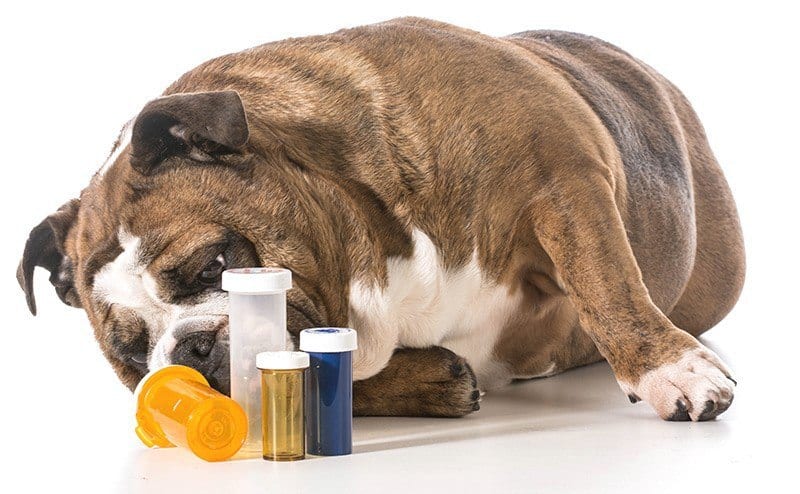
Treating Cushing's syndrome is difficult and requires close cooperation between the veterinarian and the animal's owner. Discipline in giving medication, monitoring the dog's condition and relatively frequent checkups are necessary. The dog will most often have to take medication for the rest of its life, and serious side effects fairly often accompany all forms of treatment. How effective treatment will be depends mainly on the accuracy of the diagnosis, in particular on identifying the cause, but also on treating concurrent diseases (e.g., diabetes).
It would be a truism to say that you should always be sure of the diagnosis before starting treatment. However, when such a serious disease as Cushing's syndrome is suspected, all doubts must be removed before strong and taxing drugs are introduced. The patient must have consistent clinical signs, test results consistent with the clinical course and laboratory test results confirming the suspected disease.
Sometimes Cushing's syndrome is highly likely, but the test results do not confirm it. In that case, the tests should be repeated after a few weeks, or additional tests should be considered (e.g., an ACTH stimulation test with a sex hormone panel). It also happens that a dog has minimal clinical signs while the tests confirm the disease. You should make sure the results are not false positives (this happens with severe stress or with concurrent non-adrenal illness).
There is no evidence that early treatment is beneficial, even in the early stages of Cushing's.
However, the dog should definitely be treated when the clinical signs affect the quality of life of the animal or the owner, or when there are signs that may indirectly point to HAC, for example:
- hypertension,
- urinary tract infections,
- proteinuria.
Treatment of pituitary-dependent Cushing's syndrome
Trilostane (Vetoryl) for dogs
Trilostane is the only drug registered in Poland for use in dogs with hyperadrenocorticism, and it is used as a first-line drug. Trilostane belongs to the group of reversible inhibitors of steroid synthesis. In other words, it blocks the formation of glucocorticoids and – to a lesser extent – mineralocorticoids. In this way it lowers the level of circulating cortisol, so the clinical signs subside.
The blockade of steroid synthesis is reversible, and its degree depends on the dose. Trilostane is a short-acting drug and must be given at least once a day with a meal. Most dogs tolerate it well and very rarely show signs of hypoadrenocorticism while taking it. However, a small, asymptomatic rise in blood potassium is common.
The drug is most effective in the pituitary form of hyperadrenocorticism (though not in all cases). The manufacturer recommends giving trilostane at a dose of 3–6 mg/kg body weight by mouth every 24 hours. Capsules are available in different strengths (10, 30, 60 and 120 mg). It is best given with food, as it is then most effective. The dose may be adjusted depending on the response to treatment and the results of the ACTH stimulation test.
The recommended doses are:
- 30 mg by mouth for dogs weighing <5 kg
- 60 mg for dogs of 5–30 kg
- 120 mg for dogs > 30 kg.
The effectiveness of the drug is assessed with the ACTH stimulation test, which should be performed on days 10–14, 30 and 90 of treatment, and then regularly every 3 months. The test should also be performed according to this protocol after every dose change. The blood sample should be taken 4–6 hours after the drug is given, when the drug's effect is strongest and the cortisol concentration is at its lowest – if the test is done at a different time, the cortisol concentration may have a completely different value.
- Cortisol concentration after ACTH < 20 nmol/l (0.7μg/dl) – trilostane should be stopped and the patient closely watched for signs of hypocortisolemia.
- Cortisol concentration after ACTH in the range of 0.7–1.5 μg/dl – trilostane should be withheld for 48 hours, and treatment then resumed at a dose reduced by 50%.
- Cortisol concentration after ACTH > 120 nmol/l (4.3 μg/dl) – the dose should be increased by 50–100%.
- Cortisol concentration after ACTH between 20 and 120 nmol/l (0.7–4.3 μg/dl) and the patient is doing well – the trilostane dose should stay unchanged.
If the animal is unwell (despite normal cortisol values), trilostane should be given twice a day. The clinical assessment of the patient and the ACTH stimulation test are repeated within 2 weeks of the dose change. Once the clinical signs have stabilized on trilostane, the patient is doing well and the cortisol values are within the optimal range, follow-up tests and the ACTH stimulation test should be repeated at least every 3–6 months, sometimes more often (e.g., if signs of hyperadrenocorticism or, on the contrary, of hypocortisolemia appear).
In some patients, the ACTH stimulation test results are within the optimal range, yet they still show signs of hyperadrenocorticism. In such a situation, it is recommended to split the trilostane dose into 2 doses every 12 hours, then perform an ACTH stimulation test and adjust the dose if necessary. Kidney function should be monitored with serum biochemistry and an electrolyte panel.
Many patients need trilostane dose adjustments during treatment.
Caution is needed if the patient is also receiving other drugs, such as:
- spironolactone,
- angiotensin-converting enzyme (ACE) inhibitors.
In animals with heart or kidney disease, this drug should be used very cautiously and the patient's condition checked more often, even though it is less "harmful" than mitotane (another drug used to treat Cushing's syndrome). Pregnant women should not have direct contact with it.
After the drug is started, mild side effects may appear after about 2–4 days on average, such as:
- reduced appetite,
- slight lethargy.
They often resolve on their own or after the dose is reduced.
If more serious signs appear, the drug should be stopped and the dog given prednisolone for 1–2 days. These signs include:
- diarrhea,
- vomiting,
- dullness.
Dangerous electrolyte disturbances are rare. In such situations, patients need very low doses of trilostane for the rest of their lives.
Side effects of trilostane:
- moderate drowsiness,
- decreased appetite for 2–4 days after the start of treatment (most likely due to hypocortisolemia),
- moderate electrolyte imbalances (e.g., hyperkalemia),
- rarely – signs of overt hypoadrenocorticism,
- occasional cases of acute adrenal necrosis.
If any worrying signs are noticed, the owner should contact the veterinarian as soon as possible. In such situations, it may be necessary to stop the drug temporarily, perform a stimulation test and give supportive treatment.
Mitotane
The main drug once used to treat hyperadrenocorticism was mitotane (Lysodren, o,p'-DDD). It is now successfully being replaced by trilostane. Mitotane is slightly more effective than Vetoryl but causes side effects more often. Today vets turn to it when trilostane proves ineffective. Mitotane was once used as an insecticide (a substitute for DDT), but it soon showed adrenocorticolytic properties.
It has a cytotoxic effect on the adrenal cortex, which leads to reduced production of glucocorticoids. Mitotane can cause necrosis of the entire adrenal cortex, especially the zona fasciculata and zona reticularis (the zona glomerulosa produces new adrenal cortex cells). The action of mitotane is most marked in hyperplastic adrenal glands, but its effect (a change in the stimulation test) is also seen in normal adrenal glands. That is why it must be used with great caution.
There are two options for mitotane treatment:
- Traditional, which aims to control adrenal hyperfunction in dogs without clinical signs of hypercortisolemia. It is recommended in the initial treatment of Cushing's syndrome.
- Pharmacological adrenalectomy, which aims to destroy the adrenal cortex (i.e., to go from hyper- to hypocortisolemia). It is considered only in dogs that do not respond to traditional treatment or that have stopped responding to mitotane after long-term use.
Traditional mitotane treatment consists of two stages:
- Induction (until the disease is under control).
- The maintenance phase (lifelong treatment that prevents the clinical signs from returning).
Induction phase
Before mitotane is started, the owner should measure for several days how much water the sick dog drinks and how much urine it passes per day. This is very important, because this information serves as the baseline criteria later used to assess the effectiveness of induction treatment and to decide when to move on to maintenance treatment.
Once this has been established, mitotane treatment begins:
- Treatment is started with a fairly high initial dose of 50 mg/kg body weight / day in two divided doses. Mitotane is more effective if it is given crushed, mixed with a small amount of vegetable oil and given with food.
- This treatment is continued at the recommended dose until the PU/PD clearly decreases. So the amount of water drunk and urine passed should be monitored very closely after mitotane is started as well.
- The owner should stay in constant contact with the veterinarian. Because the therapy is started at home, the dog's caregiver plays an important role in monitoring the course of treatment, in order to avoid the danger of severe hypocortisolemia developing and to move to the maintenance phase as soon as possible. The following are assessed: * the dog's activity, * level of consciousness, * appetite, * water intake, * the dog's general condition.
- Immediate contact with the veterinarian is required as soon as signs such as drowsiness, vomiting, weakness or reduced water intake are noticed. If worrying signs are found, a decision may even be made to stop mitotane.
- The average duration of this stage is 5–15 days, but large and unpredictable individual differences mean that the response to treatment can be fast and appear as early as day 3 of treatment.
- The induction phase is complete when a decrease in appetite is noted or when the dog's daily water intake falls to <80ml/kg body weight. At that point, mitotane should be stopped. This is confirmed by the result of the ACTH stimulation test, which is usually performed 5–7 days after the start of treatment. But it is the animal's caregiver who is responsible for "catching" the moment when the clinical signs of hypercortisolemia recede. That is exactly why close monitoring of the animal is extremely important.
- Failure to respond to therapy within 30 days is rare. In such a situation, it is recommended to re-examine the patient thoroughly and repeat the adrenal function tests.
- The speed with which dogs respond to the induction treatment may provide guidance for the second phase: a fast response to induction suggests a low maintenance dose, and vice versa. * The ACTH stimulation test should be performed when: * there is a noticeable decrease in appetite and thirst after mitotane is started, * the animal becomes lethargic, or diarrhea or vomiting appears, * or on day 7 of treatment. * The aim of the induction phase is slight suppression of adrenal cortex function, i.e., the cortisol level after ACTH should be in the range of 1–5 µg/dl (27–138 nmol/l).
- Induction usually takes 5–10 days. In almost all dogs with pituitary-dependent hyperadrenocorticism, a response is seen within the first 14 days of treatment. At that point, the dog should be switched to the maintenance dose of mitotane, and the ACTH stimulation test performed monthly at first, and later every 3 months.
Maintenance phase
After the test, treatment is maintained at the same dose or at half the dose once or twice a week, depending on the severity of the signs.
During therapy, the following blood levels should be monitored:
- blood urea nitrogen (BUN),
- sodium,
- potassium.
The first effects of therapy are usually seen as a reduced need for food (the dog clearly eats less and/or takes longer to finish its meal). At the same time, thirst decreases markedly – the dog drinks less than 60 ml/kg/day. During the first month of treatment, the patient becomes more alert and better able to tolerate exercise. Skin signs recede somewhat more slowly, but within 3–6 months the hair grows back normally. However, the resolution of hair loss is not linear or predictable – it largely depends on the initial degree of skin atrophy and on the time that passed between the first dermatological signs and the start of treatment.
Keep in mind that the skin condition may temporarily get worse after a few weeks of treatment. Seborrhea and itching often develop. In many dogs, the signs of the disease return after some time, and the mitotane dose has to be reviewed. If the ACTH stimulation test result suggests that adrenal cortex function has not been suppressed (cortisol concentration after ACTH > 250 nmol/l (>9 μg/dl)), the drug should be continued at the initial dose for 3 consecutive days and the effect checked by repeating the test.
It also happens that a patient develops intolerance to mitotane. Gastrointestinal signs then appear, but without a simultaneous drop in cortisol concentration in the ACTH stimulation test. These dogs should be given other drugs to treat hyperadrenocorticism.
Side effects of mitotane
- Gastrointestinal disturbances are the most common – vomiting and/or diarrhea, which force treatment to be stopped immediately or the drug to be given less often (down to once or twice a week).
- Signs of acute adrenal insufficiency (general weakness, loss of appetite, vomiting, diarrhea or coma) may appear if the therapeutic effect does not appear quickly enough or if reduced appetite or thirst is not noticed in time. Exogenous glucocorticoids may then need to be given. That is why it is a good idea for the owner to keep prednisolone at home (ideally arranged before treatment starts) so it can be given to the dog fairly quickly.
- Hyperkalemia and hyponatremia (a rise in blood potassium and a drop in blood sodium) sometimes occur. In such a situation, an ACTH stimulation test should be performed. The cortisol concentration is then usually < 20nmol/l (<0.7μg/dl). The patient needs intravenous fluids, glucocorticoids and – with hyperkalemia – mineralocorticoids. Some patients need gluco- and mineralocorticoids for the rest of their lives, while others return to a state of hyperadrenocorticism.
When switching from trilostane to mitotane (or vice versa), it is advisable to wait until the clinical signs of adrenal hyperfunction return, thus confirming the absence of adrenal suppression. However, you should not wait too long to change the drug (e.g., until the ACTH test again indicates hyperadrenocorticism), because this sometimes takes months, and serious signs of hypercortisolemia may appear during that time.
Ketoconazole
It can reduce the synthesis of steroid hormones, which is why it has been used to treat Cushing's syndrome in both animals and humans. In dogs, the classic dose is 30 mg/kg/day in two divided doses. Improvement is usually seen after 2 months of treatment. Ketoconazole has now been replaced by trilostane, and its use is controversial.
L-deprenyl (selegiline), a monoamine oxidase inhibitor
The active substance – selegiline – is used in some countries to treat the pituitary form of hyperadrenocorticism. Selegiline is a monoamine oxidase type B inhibitor (MAOBI). Monoamine oxidase is an enzyme in the brain that breaks down dopamine, so blocking it increases dopamine levels in the brain. This indirectly leads to a drop in ACTH levels (dopamine released from the hypothalamus has an inhibitory effect on the pituitary gland).
A great advantage of this form of treatment is that selegiline does not interfere with adrenal enzymes and is not adrenolytic, so monitoring is based solely on clinical signs. Hormonal monitoring is not recommended (there is no need to perform ACTH stimulation tests or measure electrolytes). It has minimal effect on the functioning of the endocrine system, which to some extent improves the patient's clinical condition.
Because of the way it works, it is used only to treat the pituitary form of Cushing's syndrome. It is given at a dose of 1 mg/kg body weight. If the clinical signs do not improve within a month, a dose of 2 mg/kg body weight/day is given. If there is no improvement within another month, treatment is stopped, because increasing the dose further will not help the patient. The effectiveness of selegiline treatment in dogs with mild or moderate signs of hypercortisolemia has been estimated at 50–70%.
In dogs with more severe signs, it is 20–30%.
Removal of the pituitary gland (hypophysectomy)
A controversial procedure that requires great surgical skill and enormous experience.
It is recommended especially for large adenomas, but postoperative complications develop very often:
frequent development of diabetes insipidus (usually transient), reduced tear production, or hypothyroidism.
Radiation therapy
It effectively reduces the size of the tumor and limits or eliminates neurological signs in dogs with signs of pituitary macroadenoma syndrome.
Treatment of adrenal-dependent Cushing's syndrome
The treatment that addresses the cause of adrenal-dependent Cushing's syndrome is surgical removal of the adrenal gland. The surgery makes sense only when we are dealing with benign, non-invasive and relatively small tumors.
The conditions for this type of surgery should be:
a highly experienced surgeon and the ability to provide proper postoperative intensive care. The controversy over adrenalectomy concerns mainly the drug management afterwards. The adrenal glands secrete not only cortisol but also other important hormones, so the fluid and electrolyte balance is severely disturbed. The matter is further complicated by the fact that the other adrenal gland is usually atrophied, so it cannot immediately take over after the opposite adrenal gland is removed.
Proper preparation of the patient for surgery can increase its chances of success. It is a good idea to start treatment with trilostane before surgery, because mitotane can make the tumor mass friable. Animals that have not been treated with drugs beforehand are difficult patients and may develop acute adrenal insufficiency after surgery. Also note that signs of hyperadrenocorticism may return even after a seemingly correctly performed procedure.
The main principles of perioperative management for adrenalectomy:
- There is no indication for giving glucocorticoids for several days before surgery, because they make fluid and electrolyte disturbances more likely and increase the risk of thromboembolism.
- At first, intravenous fluids are given (NaCl or Ringer's solution); just before surgery, dexamethasone is added to the fluids at a dose of 0.02–0.04 mg/kg body weight so that the entire dose is given over 6 hours.
- The patient should receive at least 24 hours of intensive medical care after surgery, because complications very often occur during this time.
- Blood electrolyte levels must be monitored.
- After surgery, mineralocorticoids (e.g., desoxycorticosterone acetate) are given at a dose of 0.1 mg/kg body weight.
- Dexamethasone injections are then given until the dog starts eating on its own. Once that happens, prednisone is given by mouth: an initial dose of 1 mg/kg body weight in the morning and evening for several days, then gradually reduced so that it can be stopped within 2 months.
- If sodium decreases and potassium rises to 5.5 mEq/L, mineralocorticoids must be given.
The adrenal form of hyperadrenocorticism is usually treated with drugs, using the medications listed for the treatment of the pituitary form of hypercortisolemia. Trilostane is not cytotoxic, so compared with mitotane it is probably less effective at preventing and fighting metastases.
Treatment of iatrogenic Cushing's syndrome
Long-term, intensive use of glucocorticoids suppresses ACTH secretion by the anterior lobe of the pituitary gland. Under these conditions, the adrenal cortex is not physiologically stimulated, so it secretes less cortisol, and from there it is only a step to more or less advanced atrophy. In this situation, abruptly stopping the steroid causes acute adrenal insufficiency (an adrenal crisis). That is why the drugs used so far must under no circumstances be stopped suddenly, nor their doses reduced quickly.
Signs of adrenal insufficiency include:
- agitation,
- altered consciousness,
- a weak pulse, low blood pressure,
- vomiting,
- involuntary passing of urine and feces,
- skin discoloration around the anus and vulva,
- poor appetite.
Iatrogenic Cushing's syndrome requires a completely different therapeutic approach from spontaneous hypercortisolemia, because we are in fact dealing with hypoadrenocorticism, even though the clinical signs point to hyperadrenocorticism. The corticosteroids therefore have to be withdrawn slowly.
The steroid doses should be reduced slowly over several months (3–12 months), and wherever possible the drug used so far should be replaced with another one from the same group but with a weaker action, for example:
- dexamethasone – with prednisone/prednisolone,
- prednisolone – with hydrocortisone.
Alternate-day therapy is then introduced, with the drugs given every other day. If signs of adrenal insufficiency appear, the dose should be temporarily increased or daily use resumed. If signs of adrenal insufficiency appear, the patient should be given intravenous 0.9% NaCl with added glucose and hydrocortisone at a dose of 8–20 mg/kg body weight every 4–6 hours until the signs resolve. Remember to feed a diet rich in high-quality protein.
Supportive treatment includes:
- gastroprotectants (drugs that protect the stomach lining),
- antacids,
- antisecretory drugs.
Blood potassium concentration should be monitored and any deficiency corrected if necessary.
Treating concurrent diabetes
Hypercortisolemia is often accompanied by diabetes that is difficult to stabilize despite insulin. Unfortunately, until the hypercortisolemia is stabilized, proper blood glucose control is not possible… That is why the most important goal at the start of treatment is to lower the cortisol level. Insulin treatment may be indicated in the initial period, not to achieve normoglycemia but rather to avoid serious diabetic ketoacidosis and severe hyperglycemia. That is why standard doses (0.5–1 U/kg) of an intermediate-acting insulin are recommended.
Blood and urine glucose should be monitored, but adjusting the insulin doses and achieving final blood glucose control begin only once the hypercortisolemia is under control and maintenance treatment starts. Whatever the cause of Cushing's syndrome, before deciding to start treating hypercortisolemia you should make sure the patient does not have other diseases that may affect the clinical course.
Treating hyperadrenocorticism is not without risk. The drugs used, by lowering the concentration of circulating cortisol, can cause various complications and can also worsen the signs of other concurrent diseases, such as degenerative joint disease (osteoarthritis) or atopy. There is no single, ideal way to treat hyperadrenocorticism. Given the complexity of this disease and the metabolic consequences of hypercortisolemia, each case should be analyzed in depth and the treatment tailored to the individual.
The animal's owner should be able to make an informed decision about the treatment. At the start of therapy, the least toxic drugs possible should be used, but if the patient's clinical condition does not improve within three months, the treatment should be changed without delay. Drugs that have proved ineffective should not be used either. Over time (months, years), it may be necessary to increase the dose of the drug and how often it is given.
Untreated hyperadrenocorticism
What happens if hyperadrenocorticism is not treated?
Dangerous complications may develop that – unfortunately – often end in death, such as:
- diabetes,
- systemic hypertension,
- pancreatitis,
- damage to the glomeruli of the kidneys,
- urinary stones,
- thromboembolic disease.
Cushing's syndrome: diet
This disease causes a noticeable rise in serum cholesterol and triglycerides. Excessive stimulation of lipolysis by glucocorticoids and an increased tendency to deposit fat place a heavy burden on the liver. Steroid hepatopathy may lead to bile stasis (cholestasis), which may later worsen the lipid disorders.
The diet for chronic hyperlipidemia should include:
- Feeding the dog food with a restricted fat content (<25 g/1000 kcal) and a moderate protein content (usually > 18% or 60g protein/1000 kcal).
- Omega-3 fatty acid supplementation.
- Added dietary fiber.
- Added antioxidants.
However, concurrent diseases (e.g., diabetes) should always be taken into account, so the diet should be tailored individually to each patient.
Cushing's syndrome: prognosis
The survival prognosis for dogs with the pituitary form of hyperadrenocorticism depends on the age and general condition of the animal, as well as on the owner's commitment. The average survival time from diagnosis is 30 months; in younger dogs it may be much longer (as long as 4 years). As a rule, the cause of death is complications of hypercortisolemia or other diseases of old age.
After adrenalectomy, the average survival time in dogs is about 36 months. In dogs with an adrenocortical adenoma or a non-metastasizing carcinoma, the prognosis is good. Dogs with metastatic carcinoma usually die within a year. Drug treatment of spontaneous hyperadrenocorticism makes it possible to control the signs of hypercortisolemia, but it does not cure the patient. It must continue for the rest of the animal's life, and caregivers must accept the prospect of regular checkups.
All the drugs used have side effects and certain limitations that need to be taken into account before therapy is started. Be aware that the costs of treating and monitoring patients with hyperadrenocorticism are considerable, especially during the first 6 months. In most cases, Cushing's syndrome is not a life-threatening condition for the animal, and it often affects the owner's life more than the dog's.
Sometimes treatment is limited to treating the complications associated with this disease (e.g., urinary tract infections, skin diseases or diabetes), because the dog's caregiver has limited financial means and decides not to take on the fairly high costs. At other times, the course of the disease is so mild that there are no grounds for starting treatment. Starting therapy immediately is actually necessary only in some cases, when the clinical signs are severe (e.g., marked exercise intolerance, significant obesity, diabetes that does not respond to treatment, or PU/PD so severe that the owner cannot accept it).
However, most dog owners decide to treat in order to reduce the clinical signs and the risk of later complications. In this situation, the prognosis is usually favorable. The effectiveness of treatment with trilostane has been estimated at 67% – it makes it possible to reduce the clinical signs within 3–6 months. The average survival time of dogs is 664 to 900 days. For comparison, the average survival time for dogs treated with mitotane is 810–900 days.
Summary
Finally, I would like to address the worries of owners whose dogs have been diagnosed with hyperadrenocorticism. It is undoubtedly a chronic disease, and the diagnosis itself is often hard for the caregivers of affected animals to accept. The prospect of lifelong medication, very frequent checkups and constant testing fills them with fear and sadness, as well as reluctance and doubt. In addition, the signs of overt hypercortisolemia themselves are often very hard for owners to accept.
There are situations when giving the dog away or even euthanasia is considered, because the dog urinates all over the house, sheds heavily, and its water bowl has to be refilled constantly, and so on. However, it is important to realize that this is a chronic but treatable disorder. The signs usually subside once therapy is started, and caregivers get used to regular visits and giving medication fairly quickly.
The other side of the coin is the costs, which unfortunately are quite high. The diagnostic process itself is exhausting, both in time and in money, and along the way caregivers feel irritation and doubt and put up growing resistance. Much depends here on the knowledge and experience of the veterinarian, who can skillfully choose the diagnostic tests and interpret them. Even when the owner agrees to fairly expensive tests, the result may be ambiguous and further diagnostics may be needed. This strains the patience, and also the wallet, of the sick dog's owner.
It is often very difficult to explain to caregivers that the diagnosis is also reached by ruling out other diseases, which is very important from a medical point of view but sometimes unacceptable to the owner. However, the vast majority of dog owners are capable of great sacrifices, and we vets are fully aware of this. After all, what matters most is finding the answer to the question "what is wrong with your dog" and starting the right treatment.
It will not substantially extend survival time, but it will certainly greatly improve the quality of life of both the animal and its owner.
FAQ
How long do dogs with Cushing's disease live?
The average survival time from the diagnosis of the pituitary form of hyperadrenocorticism is 30 months, and in younger dogs it may be much longer, as long as 4 years. The average survival time of dogs treated with trilostane is 664 to 900 days. The prognosis depends on the age and general condition of the animal, as well as on the owner's commitment.
What are the side effects of trilostane in dogs?
Side effects include moderate drowsiness, decreased appetite for 2–4 days after the start of treatment, moderate electrolyte imbalances, rarely signs of overt hypoadrenocorticism, and occasionally acute adrenal necrosis. If more serious signs appear — diarrhea, vomiting, dullness — the drug should be stopped and the veterinarian contacted.
How is trilostane given to a dog?
Trilostane is a short-acting drug and must be given at least once a day with a meal — it is then most effective. Its effectiveness is assessed with the ACTH stimulation test, which should be performed on days 10–14, 30 and 90 of treatment, and then regularly every 3 months. The blood sample should be taken 4–6 hours after the drug is given, when the drug's effect is strongest and the cortisol concentration is at its lowest.
Is there an alternative to trilostane for treating Cushing's in dogs?
Trilostane is the only drug registered in Poland for use in dogs with hyperadrenocorticism, and it is used as a first-line drug. The main drug used before it was mitotane. Ketoconazole has been replaced by trilostane and its use is controversial, while selegiline is used in some countries only for the pituitary form of Cushing's syndrome.
How much does it cost to treat Cushing's disease in a dog?
The costs of treating and monitoring patients with hyperadrenocorticism are considerable, especially during the first 6 months. Drug treatment makes it possible to control the signs but does not cure the patient — it must continue for the rest of the animal's life, and caregivers must accept the prospect of regular checkups.
